
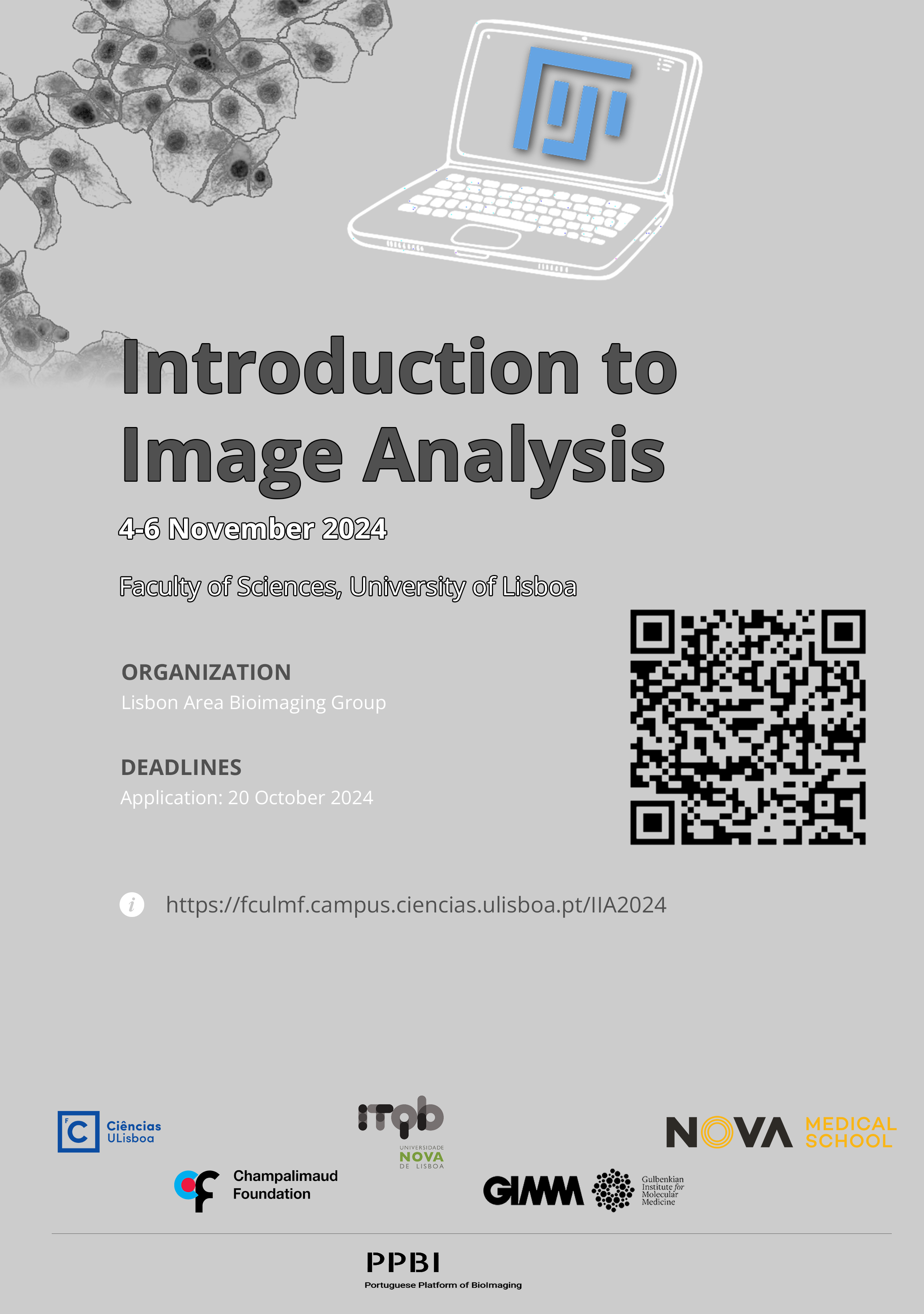
Introduction to Image Analysis – 4th edition
FCUL – 4-6 November 2024
Limited registrations: 25 participants maximum.
Overview
This course aims at providing researchers who are initiating microscopy-based projects with fundamental skills on digital imaging and biological image analysis. The course incorporates theoretical lectures as well as hands-on exercises which present key concepts and demonstrate image analysis procedures on Fiji/ImageJ, the reference open source image bioimage analysis software. Finally, trainees will learn about selecting regions of interest using artificial intelligence methods as well as recording and automating image processing workflows. Trainees will be exposed to images produced by transmission, epifluorescence and confocal microscopes. At the end of the course, trainees will be able to use Fiji/ImageJ to open, visualize and perform basic image processing and analysis procedures on their own biological images to extract quantitative information.
Program
4 November 2024
| Session 1: Working with ImageJ | |
| 9h30 – 11h00 | Fiji and ImageJ
ImageJ interface Basic concepts and common tasks Opening, visualizing and saving an image |
| 11h00 – 11h20 | Break |
| 11h20 – 12h30 | Exercises |
| 12h30 – 14h00 | Lunch |
| Session 2: Multi-dimensional images | |
| 14h00 – 15h00 | 5D images: x, y, z, channels and time
Basic operations |
| 15h00 – 15h15 | Break |
| 15h15 – 16h30 | Exercises |
5 November 2024
| Session 3: Quantitative bioimage analysis | |
| 9h30 – 11h00 | Images as a source of data
Regions of interest Line profile ROI Manager |
| 11h00 – 11h20 | Break |
| 11h20 – 12h30 | Exercises |
| 12h30 – 14h00 | Lunch |
| Session 4: Spatial measurements in ImageJ | |
| 14h00 – 15h00 | Pixel spatial calibration
Setting a scale bar Measurement types |
| 15h00 – 15h15 | Break |
| 15h15 – 16h30 | Exercises |
6 November 2024
| Session 5: Image pre-processing and segmentation | |
| 9h30 – 11h15 | Basics of linear filtering
Thresholding Basics of Binary filtering Object counting |
| 11h15 – 11h30 | Break |
| 11h30 – 12h30 | Exercises |
| 12h30 – 14h00 | Lunch |
| Session 6: Advanced Segmentation | |
| 14h00 – 15h00 | Labkit
Stardist |
| 15h00 – 15h15 | Break |
| Session 7: Introduction to automation | |
| 15h15 – 16h15 | Using the macro recorder
Introduction to batch processing Best practices in reporting bioimage quantification |
| 16h15 – 17h00 | Exercises |
Faculty
Venue
The course will take place at FCUL from 4th to 6th of November 2024.
We will be meeting at 4.2.07 (Building C4, 2nd Floor, room number 7). This is within the main library building. There are multiple options for meals inside and outside the FCUL campus.
FCUL is located next to Campo Grande and Cidade Universitária. We recommend traveling by public transportation. There is parking for private vehicles, but spots are always in high demand.
Partners


Poster
Contact
Hugo Botelho
Faculty of Sciences, University of Lisboa