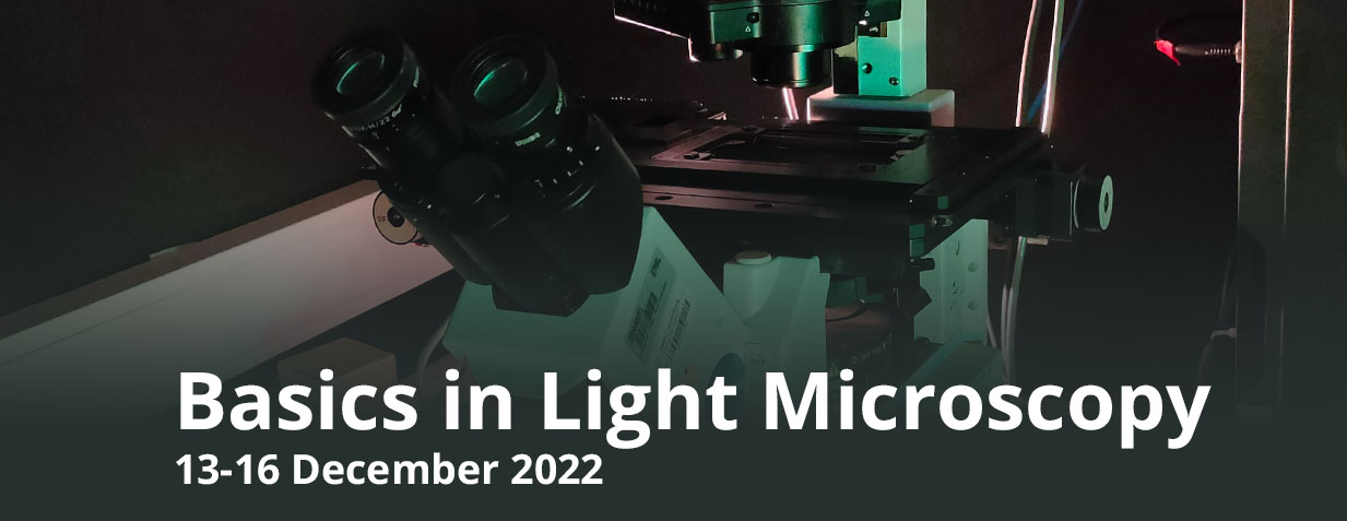
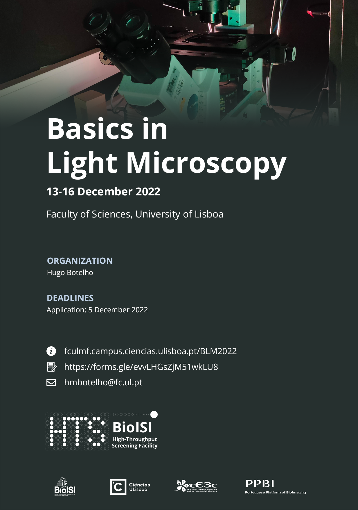
FCUL Course – Basics in Light Microscopy
Date: 13-16 Dec 2022
Time: 9h30 – 16h00
Description
This course will provide a solid understanding of the fundamental principles of light microscopy for cellular and developmental biology researchers. Through a series of interactive tutorials and hands-on sessions, trainees will be exposed to the photophysical, spectroscopic and optical background to optical microscopy, as well as instrumental and biological sample preparation considerations. Emphasis will be placed on bioimaging technologies available at FCUL: transmission, epifluorescence and confocal fluorescence microscopies. At the end of the course a short overview of advanced light microscopy technologies will be presented. This course is targeted to young researchers who use or intend to use light microscopy as part of research projects, especially MsC, PhD students and post-docs who did not have formal introduction to microscopy as part of their academic training. Attendance will be limited to FCUL members.
Poster
Target audience
FCUL Members; MsC and PhD students. Post-docs and facility trainees.
Cost
Free.
Format
In-person course.
Slots: 15 attendees
Registration
Application deadline: 5 December 2022
Acceptance notification: 6 December 2022
Registration form must be submitted along with a motivation statement.
Trainers
- Hugo Botelho
- Luís Marques
Program
- Introduction
-
- Electromagnetic nature of light (frequency, wavelength, polarity, phase, speed)
- Refraction and reflection
- Jablonski diagram: Absorption and fluorescence
- Timescales of photophysical processes
- Photobleaching
- Limitations in optical microscopy
-
- Chromatic aberration
- Spherical aberration
- Field curvature
- Absorption Dyes
-
- Some common dyes: Hematoxylin & eosin, Giemsa, Gram, Propidium iodide
- Fluorescent Dyes
-
- Major fluorophore families (xanthene, indole, cyanine, alexa)
- Fluorescent proteins
- Quantum dots
- Assay considerations
-
- Fixation
- Immunofluorescence
- Organelle-specific dyes
- Autofluorescence
- Mounting media
- Live cell imaging
- Basic aspects of keeping cells alive during an observation
- Bringing live cells into the microscope stage
- The compound optical microscope
-
- Schematic optical path
- Conjugated planes
- Infinity corrected optics
- Optical components
- Light sources
- Filament bulb (e.g. metal halide)
- LEDs
- Arc Lamp
- Lasers
- Single wavelength and super-continuum (WLL)
- CW vs pulsed
- Objectives
- Magnification
- Numerical aperture
- Chromatic correction
- Immersion
- Eyepiece
- Filters and bleedthrough
- Dichroic mirrors
- Array detectors (CCD, CMOS). Black & white and color.
- Point detectors (PMT, GaAsP, APD, Hybrid)
- Spectral detection
- Detector linearity
- Light sources
- Slide and coverslip
- The image of a point is not a point
- Resolution and convolution
- Bright field microscopy
-
- Introduction – what it is
- Kohler illumination
- Phase contrast
- DIC
- Dark field
- Epifluorescence microscopy
-
- Introduction – what it is
- Optical setup
- Filters and filter cubes
- Optical sectioning microscopy
-
- Introduction – what it is
- Principles of the confocal microscope
- Laser boxes
- Pinhole and confocality
- Comparison with epifluorescence – differences in construction (same presentation)
- Noise, resolution,
- Spinning disk confocal
- Advanced techniques
-
- Multi-photon confocal
- Optical sectioning: light sheet
- High-throughput microscopy
- F techniques: FRAP, FRET, FLIM, FCS, FCCS
- Super-resolution
- TIRF
- SRRF
- STED
- PALM/STORM
- Structured illumination
- Digital image basics
-
- An image as a matrix
- Bit depths
- pixels/voxels
- 5D images
- Image formats
- Compression: lossy/lossless
Timetable
| Tue 13 December |
Wed 14 December |
Thu 15 December |
Fri 16 December |
|
| 9h30 – 11h10 | Presentation Intro: Absorb. and Fluor. Aberrations |
Compound optical microscope (I) | Brightfield Epifluorescence | Advanced techniques |
| 11h10 – 11h30 | Coffee Break | Coffee Break | Coffee Break | Coffee Break |
| 11h30 – 13h00 | Fluorescent dyes Labeling and assays |
Compound optical microscope (II) | Confocal | Digital image basics |
| 13h00 – 14h30 | Lunch | Lunch | Lunch | |
| 14h30 – 16h00 | Practical: brightfield microscopy | Practical: widefield fluorescence microscopy | Practical: confocal microscopy |
Contacts
htsfacility@fc.ul.pt
Hugo Botelho